

Digital Microscopy in 2026: Cameras, Software & Imaging Integration
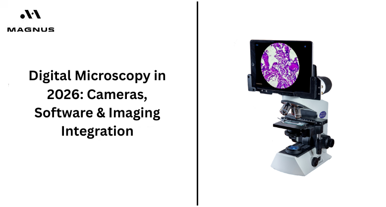
In the world of advanced technologies, the introduction of digital microscopes in 2026 gives an edge in the industry. It helps users to go far beyond simple magnification and get scientific images with the advanced cameras, AI software and connected imaging systems.
The digital microscopy sensors capture minute detail, while AI-enabled software automates image analysis, measurement, and defect detection with accuracy. These advanced tools help laboratories, healthcare facilities, and manufacturers to improve efficiency, reduce errors, and accelerate research outcomes. Its seamless integration of cloud technologies and digital databases also allows users to manage and share imaging data easily across teams.
Let's dive into the blog to explore how digital microscopy in 2026 helps users in their field with its clarity camera pictures and software integration.
What Makes The Digital Microscope Special in 2026
In 2026, digital microscopy will change a lot in the workspace. Despite the magnification lenses, it now uses AI tools and high-quality integration to capture accurate images that are beyond what the eye can witness. Many researchers in labs, factories and schools are using digital microscopes like MX21i to capture clear and detailed studies of the sample.
The advanced software makes the image analysis easy, while the integration connects everything smoothly. It gives a live video analysis and a 3D image-capturing tool, which means users can gain instant updates of the sample and spot problems quickly. Its quick functionality minimises the work time in the lab and lets you learn more. It helps users to be more flexible with the technology at a low cost.
What Makes a Digital Microscope Camera Stand Out?
In digital microscopy, camera magnification is what rules the system. Its CMOS sensor captures images with 4k clarity and gives a standard analysis with zero errors. Its DSX pack has 12MP for all lights, and the TOMLOV DM9 has a 7-inch screen and autofocus images to capture sharp images.
As the digital microscope camera comes with the AI integration of HDMI and USB, users can gain instant analysis records of the sample and reduce errors by 31%. The MX21 digital microscope uses a 5MP USB 2.0 or MagCam up to 10MP for fast functionality.
With its advanced lens, users can capture images 49x to 1000x, where its infinity optics keep the view flat and bright. Whether you are using it to study cells or field work particles, its camera ensures the capture of the images in detail under your budget limit and handles daily use.
Software: Digital Ready Workstation
The digital microscope is an optical workhorse equipment which has a parfocal, achromatic objective train and coaxial coarse. Its fine focus and robust mechanical stage help users to analyse the image closely without pixelisation. It relies completely on a vendor-supplied imaging software microscope system that can capture, process and manage images from the connected cameras. Along with giving accurate measurements, it performs image enhancement and annotation for smooth working.
- Its high-quality camera captures a live-view image in detail and measurement on the connected 5 to 8 MP stream.
- The annotation and teaching tools in digital microscopy let instructors mark the cell structure and label the regions of interest. It saves images for students so that they can use them for revision and as exam material.
- With its data storage software, users can revisit the captured image or marked lesson with metadata for later reference. Its flexible usage allows schools and clinics to take a low‑cost MX21i microscope and upgrade its software layer to match evolving digital‑microscopy trends without replacing the core optics.
How Does Digital Microscope Imaging Integration Work?
The digital microscope is built with ergonomic and molecular design, which makes it easy to integrate into varied imaging workflows. Its LCD screen replaced the traditional eyepiece with a 10.2-inch screen and an 8MP camera for better capturing and image analysis. It gives
- Direct viewing and immediate capture without needing a separate monitor or PC.
- With standard Wi-Fi connectivity and onboard storage, the image change can be saved and shared with the lab computer or drive easily.
- For example, in teaching labs, the instructor can project live images from an LCD microscope so the students can learn through structure and export annotated images for study material.
- In clinical and diagnostic setups, it supports slide documentations, ensuring attachment-related images on patient records and morphological measurements without complexities.
Traditional vs Modern Digital Microscopes in 2026
In 2026, the cutting edge of digital microscopy is defined by various features.
- Along with giving 4K clearness, the digital microscopy gives a higher CMOS camera with USB3 vision. Its HDMI output and high frame rates support users in capturing accurate imaging without any blurry edges.
- The AI-driven tools help in giving super-resolution performance, defect classification and cell segmentation automatically without any delays.
- Integrated digital microscopes give users onboard analysis, robotic stages, and cloud connectivity.
By these standards, the digital microscope acts as a mid-range and analogue core instrument that becomes digital through its camera and software add-ons. It lacks automated XY stages, motorised Z‑drives, and high‑speed, high‑resolution cameras.
- Compared to other microscopes, it comes with a lower cost of entry for schools, small clinics and rural labs.
- Its robust optics with an achromat objective and stable mechanical stage give suitable options for thousands of slide changes.
- Its digital hybrid operation helps users to view samples through the eyepiece while training students or patients on a digital screen.
How to Choose the Best Digital Microscope in 2026?
Here are the practical tips that users can follow when choosing a digital microscope.
- Choose the right camera configuration for image capturing. For basic documents, they can have a 5MP USB connector camera, while an 8MP camera is for detailed image capturing.
- Use standard image software that comes with live view, measurement and annotation, and easily export the documents into a common format.
- It helps to simplify the work and gives standard guidance for students treating patients.
- Use a centralised folder or simple database to store images with metadata.
Final Takeaway
In 2026, digital microscopes come with high resolution, AI-infused software and well-connected integration for a smooth workflow from hardware to cloud platform. If you are looking for a budget-friendly yet advanced software microscope, then check with Magnus Opto, which offers a leading-edge supply of premium digital microscopy within your budget.
Magnus Opto offers a cost-effective solution with an optically robust microscope that can be turned into a digital-ready workstation. Along with quality manufacturing, we also provide reliable delivery and installation guidance for every user.
Contact Magnus Opto now to book your digital microscope!
FAQs
Q1. What is a digital microscope and how does it work?
A digital microscope uses a camera sensor to capture magnified images and display them on a computer, monitor, or other digital device. Modern systems can combine cameras with imaging software for measurement, annotation, image processing, and documentation.
Q2. What microscope camera resolution is best for digital microscopy?
The ideal resolution depends on the application. A 5MP camera can work well for routine documentation and teaching, while 8MP or higher cameras are better suited for detailed imaging, research, and image analysis.
Q3. What software is used with a digital microscope camera?
Digital microscope software can provide live viewing, image capture, measurement, annotation, enhancement, and data management. Popular microscopy software options in 2026 include ImageJ, Fiji, and Micro-Manager, depending on the camera and workflow.
Q4. Can AI be used for digital microscope image analysis?
Yes. AI-assisted microscopy can help with tasks such as cell segmentation, object detection, classification, counting, and image analysis. AI is increasingly being integrated into microscopy workflows to improve efficiency and support researchers and laboratory professionals.
Q5. How do I connect a microscope camera to a computer?
Most digital microscope cameras connect through USB, while some systems also support HDMI, Wi-Fi, or dedicated imaging platforms. Before purchasing, check the camera's software, operating-system, interface, and microscope compatibility.
Q6. What should I look for when buying a digital microscope in 2026?
Consider camera resolution, frame rate, sensor quality, microscope compatibility, software features, connectivity, image storage, measurement tools, and upgrade options. For routine laboratory use, a reliable optical system combined with a suitable camera and imaging software can provide a practical digital workflow.