

Trinocular Microscopes Explained: Why Labs Choose Digital Imaging Setups
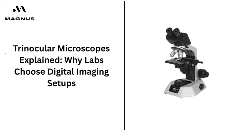
In today's laboratory environments, there is a need for precision, efficiency, and accurate observation documentation. It is for this reason that trinocular microscopes have become important equipment in learning institutions, pathology laboratories, research centers, and industries. Contrary to other conventional microscopes, trinoculars offer an extra eyepiece where one can connect an image device.
With the growing adoption of digitization in modern laboratories, microscopes with advanced imaging capabilities are highly preferred. Scientists, instructors, and lab technicians need equipment with high optical quality but with digital features that make their work easier and more efficient. The presence of a digital microscope camera allows individuals to record videos and take images.
The following blog highlights reasons why laboratories are embracing trinocular microscopes, their benefits, and digital imaging advantages in scientific fields.
Definition of a Trinocular Microscope
A trinocular microscope denotes a sophisticated version of a light microscope that has three openings for viewing. While two viewing ports are used to look through, the third opening is meant for cameras and similar devices.
Main parts of Trinocular Microscopes:
1. Binocular Lenses
- This piece of equipment enables users to see specimens in microscopes.
- Without straining their eyes.
2. Third Viewing Opening
- This part becomes handy when attaching camera systems to your trinocular microscope.
3. Magnifying Objectives
- Several objective lenses have been invented, including lenses of 4x, 10x, 40x, and 100x.
4. Light Sources
- LED and halogen lights improve the viewing process.
5. Mechanical Stage
- Microscope stages enable users to move and position slides on the microscope.
It is the third viewing opening that makes these microscopes useful in modern digital laboratories.
Reasons Laboratories Favor Digital Imaging Systems
Digital imaging has revolutionized laboratory procedures in various industries. In biology, medicine, pharmaceutical, or material science, laboratories have gained advantages from recording their visuals digitally.
1. Better Documentation
Better documentation is one of the primary reasons laboratories opt for digital imaging systems.
Advantages include:
- Recording microscopic objects in real-time.
- Storing images for later use.
- Generating comprehensive laboratory reports.
- Facilitating the easy dissemination of information to other teams.
A microscope with a camera makes it easier for scientists to keep proper documentation.
2. Improved Cooperation and Training
The digital image allows more than one user to view the specimen at once.
Advantages for educational and research laboratories:
- The images can be shown on computer screens or through projectors.
- Viewing of demonstrations becomes easier.
- People can share their research findings with others.
It especially helps in educational facilities where the specimens need to be explained to several people.
3. High-Definition Image Capturing
Modern microscopes are equipped with imaging systems that facilitate the production of high-quality visual images.
Characteristics of Imaging System:
- Capture of high-definition images.
- Video capability for recording motion in the specimen.
- Ability to zoom and measure.
- Software support for image enhancement.
The digital microscope camera offers laboratories a means of capturing accurate visual images.
Usage in Medical and Pathology Labs
A trinocular microscope is highly important in today's medical diagnostic and laboratory setups. The capability of the trinocular microscope to perform digital imaging proves its usefulness in diagnostics.
Examination of Blood Sample
The use of a trinocular microscope helps the medical laboratory personnel analyze blood samples, as well as identify any abnormalities that may exist.
Examining Tissue
A pathology laboratory can examine tissue using a trinocular microscope.
Examination of Cell Structure
Scientists can employ a trinocular microscope in order to study cell structure.
Diagnosis of Diseases
Disease can be efficiently diagnosed by capturing images through a trinocular microscope.
Educational Institutions
Schools and colleges utilize high-tech microscopes for enhanced practical education.
Advantages of Education:
- Students gain deeper insight into specimen characteristics.
- Images can be stored and reused by instructors for demonstrations.
- Training centres can maintain electronic records of their collections.
The use of a microscope with a camera aids in closing the gap between conventional microscopy and educational technology.
Industrial and Quality Control Labs
Industries rely on microscopes for inspection and defect analysis.
Common uses:
- Semiconductor inspection.
- Textile fiber analysis.
- Material surface evaluation.
- Electronics testing.
Digital systems help technicians document defects and maintain quality assurance records.
Benefits of Utilizing Trinocular Microscopes
Increased Efficiency within Laboratories
Labs can benefit from the use of trinocular microscopes due to the fact that they enable several people to observe and record images under the microscope at once.
Proper Archiving
Thanks to the fact that the images can be saved and later accessed, it is much easier to keep the necessary data for various uses in the laboratory or educational process.
Facilitation of Communication Processes
Images taken using such devices can easily be transferred to other scientists, students, or physicians.
Importance of Digital Imaging in Research
Digital imaging is an important tool for researchers because it allows them to take photographs for observation and documentation. Digital imaging also enables them to store the photos for later comparison.
Scientists are therefore able to analyze the results from experiments more accurately, and their work becomes reliable. The process also minimizes errors that may occur when doing manual observation and helps to improve how they present their results.
Digital imaging also facilitates collaboration between scientists by making image sharing easy. It also allows real-time analysis and advanced research through the application of digital imaging in medicine, microbiology, biotechnology, and material testing laboratories.
The Reason for Digital Configurations Being the Future of the Lab
Labs are adopting a paperless environment. An imaging system will aid this process by enhancing efficiency and precision.
The Increasing Role of Imaging Technology:
Contemporary scientific research demands:
- Precision in digital record-keeping.
- Speed in communication.
- Enhanced visualization technologies.
- Effective data management.
An ideal solution would be a trinocular microscope because it integrates perfectly with the current lab requirements.
Final Thoughts
The trinocular microscope has proven itself indispensable in laboratory settings today due to its optical accuracy and digital imaging abilities. For those who are on the hunt for quality microscopy services, Magnus can be counted upon as an industry expert.
Magnus has earned a reputation as a producer of sturdy and precision-oriented microscopes suitable for educational, diagnostic, and research purposes. The company also provides dependable customer support, installation assistance, product guidance, and reliable after-sales services for laboratories and institutions.
Frequently Asked Questions
Q1. What is a trinocular microscope and how does it work?
A trinocular microscope has three optical ports: two for viewing through the eyepieces and a third for connecting a microscope camera. This allows users to observe specimens directly while simultaneously capturing images or videos for digital documentation.
Q2. What is a trinocular microscope used for?
Trinocular microscopes are used for medical diagnostics, pathology, biological research, education, microbiology, and industrial inspection. Their camera port makes them particularly useful when microscopic images need to be captured, analyzed, stored, or shared.
Q3. What are the benefits of using a digital camera with a trinocular microscope?
A digital camera enables high-resolution image capture, video recording, digital documentation, image sharing, measurement, and annotation. It also allows multiple users to view the specimen on a computer or display instead of relying only on the microscope eyepieces.
Q4. Can a trinocular microscope be used for pathology and medical diagnostics?
Yes. Trinocular microscopes can support blood smear examination, tissue observation, cell analysis, and pathology documentation. When connected to suitable imaging software and a microscope camera, they can also help laboratories create and share digital records.
Q5. What is the difference between binocular and trinocular microscopes?
A binocular microscope has two viewing ports for direct observation, while a trinocular microscope adds a third port for a camera or imaging device. The trinocular configuration is better suited to laboratories that need both direct observation and digital image capture.
Q6. How do I choose a trinocular microscope for digital imaging?
Consider optical quality, objective magnification, camera compatibility, illumination, mechanical stage, imaging software, camera resolution, and connectivity. For research, diagnostic, or educational use, choose a system that supports the required image capture, measurement, documentation, and sharing workflow.