

Microscope Calibration Guide: Stage vs Eyepiece Micrometre
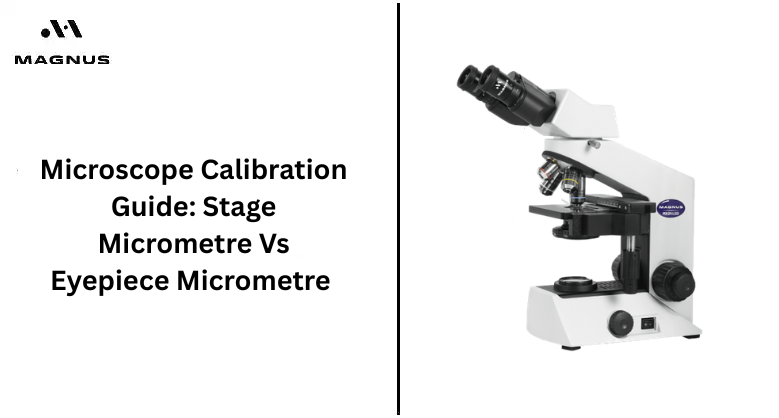
Accurate measurement plays an important role in laboratory observation, especially while examining biological samples, industrial materials, or research specimens under different magnifications. A proper microscope calibration process helps users understand the exact size of the structures they observe through the microscope lens.
Many professionals depend on calibration methods to improve consistency during routine analysis and educational work. The difference between a stage micrometre and an eyepiece reticle creates a stronger foundation for precise microscopic measurement in laboratories, research centres, and teaching environments.
Role Of A Stage Micrometre
A stage micrometre is a specially marked glass slide that contains a fixed measurement scale etched with high precision for microscope calibration procedures. Most stage micrometres contain one millimetre divided into one hundred equal sections, where each division represents ten micrometres under standard measurement conditions.
The slide is placed directly on the microscope stage during calibration work. Its fixed scale acts as a known reference that helps users determine the actual value represented by divisions inside the eyepiece reticle. The stage micrometer and eyepiece remain constant regardless of magnification changes, which makes it an essential tool for accurate measurement comparison.
Working Principle Of An Eyepiece Micrometre
An eyepiece Micrometer contains a small glass disk installed inside the microscope eyepiece, displaying evenly spaced divisions that help estimate specimen dimensions during observation. The scale itself does not contain fixed measurement values before calibration takes place under a selected objective lens.
The apparent size of the reticle changes according to the magnification levels used during microscope operation. A division observed under low magnification represents a different measurement value when viewed under higher magnification settings. This changing relationship creates the need for proper calibration before measurement work begins in laboratory environments requiring dependable results.
Calibration Practices For Consistent Laboratory Results
Accurate Calibration supports reliable specimen measurement in microbiology, pathology, material science, educational research, and industrial quality inspection activities performed under laboratory microscopes. Even small calculation errors may influence analytical conclusions during projects involving cell dimensions, particle analysis, or structure comparison studies.
Consistent calibration practices reduce the risk of measurement variation between different microscope users working within the same laboratory department. Proper alignment procedures improve observation confidence while supporting better record-keeping during scientific documentation tasks. Careful calibration habits contribute to long-term microscope maintenance and improve the overall performance of optical systems used for precision-focused work.
Step-By-Step Calibration Procedure
The calibration process begins by inserting the reticle into the microscope eyepiece and adjusting focus until the scale appears clear during observation.
The stage micrometer is then positioned on the microscope stage and brought into sharp focus using a lower-power objective lens.
The next step involves aligning both scales until they appear parallel inside the field of view. Users align the zero marking on each scale, then identify another matching point farther along the measurement line. The known stage micrometer distance is divided by the number of eyepiece divisions between matching points. This calculation provides the value represented by one reticle division under that specific objective lens.
Simple Comparison Between Both Micrometres
The stage micrometre and eyepiece reticle perform connected roles during calibration, though each tool serves a different purpose within the overall measurement process. One acts as the standard reference scale, while the other becomes the measuring scale after calibration is completed correctly.
| Feature | Stage Micrometre | Eyepiece Micrometre |
|---|---|---|
| Position | Placed on the microscope stage | Installed inside the eyepiece |
| Scale Type | Fixed measurement scale | Arbitrary division scale |
| Main Purpose | Reference for calibration | Measurement after calibration |
| Magnification Effect | Remains constant | Changes with magnification |
| Usage Frequency | Used during calibration | Used during specimen measurement |
These differences help users avoid confusion during routine laboratory work. Clear identification of each component improves efficiency during calibration sessions involving multiple objective lenses.
Why Must Calibration Be Repeated For Each Objective?
Many microscope users assume one calibration setting works for every magnification level, though the relationship between scales changes whenever the objective lens changes. The visible distance covered by the stage scale becomes smaller under higher magnification, creating a different comparison result with the eyepiece divisions.
A calibration value obtained under a 4x objective cannot provide accurate measurements under 10x, 40x or 100x objectives. Each lens requires a separate calibration record for dependable specimen analysis. Laboratories often maintain written calibration charts near workstations, allowing users to quickly reference measurement values during routine examination procedures involving multiple magnification settings.
Common Errors During Calibration
Several common mistakes reduce measurement accuracy during calibration sessions, especially when users rush through alignment procedures without carefully checking scale positions. Non-parallel scales create inconsistent readings that affect calculations and produce unreliable specimen measurements during laboratory observations.
Improper focusing creates blurred division that makes matching points difficult to identify under the microscope's field of view. Some users forget to recalibrate after changing objective lenses, leading to incorrect measurements during detailed analysis work. Dust accumulation on optical components may reduce image clarity during calibration sessions. Regular cleaning routines, careful handling practices and a stable microscope positioning improve measurement consistency during daily laboratory operations.
How To Improve Calibration Accuracy?
Users can improve calibration quality by beginning with lower magnification objectives before moving toward higher-power lenses requiring finer alignment precision. Careful focusing helps create sharp scale marking that supports easier identification of matching divisions during calibration calculations.
Stable lighting conditions improve visibility while reducing strain during extended microscope sessions involving repeated measurement work. Calibration records should be updated regularly whenever optical components are serviced or replaced within the microscope system. Laboratory staff benefit from standardised calibration procedures that maintain consistency across multiple instruments.
Clear documentation practices support better measurement reliability during educational training, scientific studies, and industrial quality-control applications requiring dependable microscopic analysis.
Final Thought
Accurate microscope measurement depends on how the stage micrometres and eyepiece micrometre scale work together during laboratory observation procedures. Reliable microscope calibration supports precise specimen analysis and dependable measurement consistency under different objective lenses used during scientific work.
The stage micrometre provides the fixed reference needed for calculating measurement values, while the eyepiece micrometre becomes useful after proper calibration is completed for each magnification level. Laboratories seeking high-quality optical solutions and dependable calibration support the products and guidelines available in Magnus Opto for professional microscopy applications and requirements.