Phase Contrast Microscopy: Visualizing Unstained Samples with Magnus Optics
Scientists often require direct observation of living cells to understand their function and processes. The cells are translucent and hardly visible under a typical microscope. It is quite a challenge to examine their morphology and interior components.
Phase contrast microscopy is a technique that is highly useful in such situations. It is a unique method that permits the visualization of transparent specimens and images. Making it possible to observe them without the need for dyes or stains. A phase contrast microscope changes very small changes in light into clear contrast.
Because of this change, the inside parts of cells become easier to see. This method is used in many biology, research, and medical labs. It helps scientists with cells that are still alive. Studying living cells is important because it shows how cells behave in their natural state.
Why Transparent Cells Are Hard to See
Living cells are completely transparent. When light passes through them, the brightness changes very little. A normal microscope shows objects mainly based on how they block or absorb light. These cells do not block the high-volume light.
They look almost invisible, very faint. Scientists frequently apply stains to colour cells to fix this. They make the nucleus and the cell membrane visible. Stains can harm live cells or change their behavior. It becomes very difficult to study cells in their natural condition. Phase contrast microscopy is one way to visualize cells without staining.
The Basic Idea of Phase Contrast Microscopy
The main principle of phase contrast microscopy is the property of light to change its phase. Its speed as it travels through different materials. When light enters various regions of a cell, it is transmitted to varying degrees depending on the density of that region. This slight difference results in a phase shift of the light waves.
Human eyes are not capable of detecting such minute phase differences. A phase contrast microscope transforms these hidden phase variations into visible intensity contrasts. Certain cell components look darker and others lighter. This greatly facilitates the visualization of the cell's architecture.
How the Technique Was Developed
Phase contrast microscopy was a concept developed by a scientist. In the first decades of the 20th century, Frits Zernike stumbled upon this phase, and his finding completely transformed how scientists investigate cells. They relied on stains to make different parts of cells visible. These stains sometimes harm cells or alter their natural state.
Zernike's method allowed scientists to observe living cells without using stains. This made it possible to observe cellular actions such as growth, movement, and division as they occur. Today, this method is still widely used for analyzing unstained cells in many laboratories.
Important Parts of a Phase Contrast Microscope
A phase contrast microscope has special optical parts that help create clear images. Two important parts are the condenser annulus and the phase plate inside the objective lens. A ring of light shines on the sample. Some light goes straight.
A bit changes because of cell parts. The phase plate makes those small shifts visible. If everything lines up right, the microscope shows a clear picture. Seems like the setup helps see details better. Plus, the changes are subtle but noticeable.
How Phase Contrast Imaging Works
The process of phase contrast microscopy begins when light passes through the condenser and reaches the sample. As light moves through the specimen, some light passes straight through, while other light bends when it encounters cell structures.
Both types of light then move into the objective lens. Inside the lens, the phase ring changes the light that passes directly through the sample. When straight and bent light combine, they create differences in brightness. These brightness differences form the final image seen in the microscope. Even the clear parts of the cell become visible.
Benefits of Observing Unstained Samples
The benefit of phase contrast microscopy is that it allows scientists to study cells without using stains. This is very useful when working with living samples.
Using an Inverted Microscope, the cells remain alive and behave naturally. The benefit is that samples can be observed for longer periods without damage. These advantages make unstained cell analysis widely used in many areas of biological research.
Uses in Scientific Research
Phase contrast microscopy works in many scientific fields. The cell biology track covers cell division, movement, and internal changes. Labs use it to check cultured cells and biological samples. It's useful for biotech and medical research.
When making new medicines, scientists need to watch how cells react. This microscope helps them do that. Probably the best tool for seeing tiny life in motion. It seems widely trusted for live-cell views.
Limitations of Phase Contrast Microscopy
The phase contrast microscopy is very useful. It has some limitations. The common issue is the halo effect that sometimes appears around the edges of objects. These bright halos can hide small details in the image.
Another limitation is that thick samples may not produce very clear images. This method works best with thin and transparent samples. These limitations of the phase contrast microscope remain a useful tool for studying living cells.
Future of Phase Contrast Imaging
Microscope technology continues to improve due to new techniques. The advancements in digital tools and sharper lenses. Today's models mix phase contrast with smart cameras and software. This helps researchers see clearer pictures and examine cell details more closely.
Plus, it seems like the results are getting much more detailed over time. Digital systems and magvision software also make it easier to store, share, and study images. As technology continues to improve, phase contrast imaging will remain very important for studying living cells and performing detailed, unstained cell analysis.
Winding It Up
Seeing clear shapes in invisible parts of life became possible through phase-contrast techniques with light. Instead of dyes, tiny brightness shifts reveal what lies inside living cells. While regular scopes fail, this version captures fine inner layouts without harm.
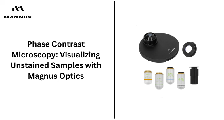
Alive and natural, specimens show their true form over time. Because precision matters, Magnus Opto builds high-quality lenses tuned for real-world labs. With sharp lenses and straightforward settings, their microscopes deliver clean images and precise viewing.
When clearer cell details matter, looking into what Magnus Opto offers might just make sense. A lab aiming for stronger outcomes could find these tools to be a good fit. Moving ahead often means choosing gear that simply works.
With advanced features that encourage collaboration and deepen scientific understanding, Magnus Opto equips users with the tools they need to explore and learn. Choose Magnus Opto to bring the fascinating world of science closer!