Top Research Microscopes in Hyderabad for Modern Scientific Studies
Hyderabad's state-of-the-art imaging facilities and instrument labs have made it a major centre for scientific research and cutting-edge microscopy. Nowadays, facilities provide a variety of microscopes, such as confocal, super-resolution, cryo-electron, and scanning electron microscopes, which are essential for contemporary research in biology, chemistry, materials science, and the environment. Scientists can examine cells and materials in more detail due to advanced microscopes, which advance research in important areas like biology and nano. Learn about the top research microscopes in Hyderabad as you read this post until the end.
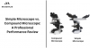
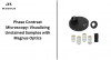
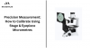
Phase Contrast Microscopes
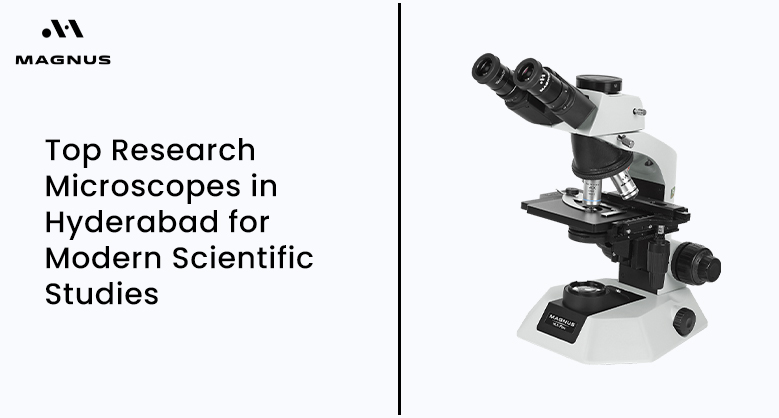
Transparent and unstained cells are easier to see with phase contrast microscopes, one of the popular research microscopes in Hyderabad. For the study of live cells, including sperm analysis, they are perfect. Magnus provides these characteristics in their MLXi Plus series. For improved picture clarity, the microscopes use phase coatings created in Japan. There exist trinocular and binocular models. These include a universal condenser, wide-field eyepieces, and a mechanical stage.
The lighting options include "Freedom" and LED variants. High resolution and ease of use characterize these microscopes. Labs that need to examine living, moving cells without staining them are ideal.
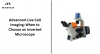
Confocal & Super‑Resolution Microscopes
Super-resolution and high-end confocal systems are now used in Hyderabad research labs. One example is the ECLIPSE Ti2 inverted confocal system at the Nikon Centre of Excellence at IIT Hyderabad, which has two scanners and super-resolution capabilities. Being one of the best research microscopes in Hyderabad, it enables live-cell imaging using CO₂ incubation and TIRF.
This device can capture high-quality photographs of subcellular structures and tagged biomolecules.
IIT Hyderabad's SATHI facility has a Nikon AXR Ti2E confocal with a Nikon Spatial Array detector. The lateral resolution it provides is 100 nm. Additionally, it supports DIC, FRET, FRAP, FLIM, and TIRF. It allows 0.01 µm steps for z-stacking and image stitching. Features for live cell incubation are included. These capabilities aid in a more thorough analysis of cellular activity.
A spectral confocal microscope A1R si with resonance scanner at RGCB can capture images at 512 x 512 resolution at over 25 frames per second. Ar, diode, and He-Ne lasers and a 32-array spectral detector are used. Imaging at 100 nm resolution is made possible using a specialized super-resolution N-SIM module. This allows both live-cell and fixed-cell imaging processes. The Leica SP8 WLL confocal at RGCB also offers GaAsP detectors and tunable lasers for intricate multi-fluorescence studies.
Stereo & Wide‑Field Fluorescence Microscopes
Stereo fluorescence microscopes are necessary for rapid sample preparation and inspection. Additionally, Nikon CoE has stereo fluorescence devices. GFP filters provide wide-field imaging.
Whole-organism imaging and tissue sections can benefit from these methods. Additionally, Leica's lineup includes modular stereo fluorescence systems. These devices form an important part of the collection of research microscopes in Hyderabad, especially for life science studies and specimen-level analysis.
Scanning Probe and Electron Microscopes
Hyderabad labs offer electron microscopy in addition to optical imaging. Icon Analytical lists Hitachi SEM systems with cooled stages for samples sensitive to beams. Electron microscopes and training support are provided by Beeta Tech India, a company situated in Hyderabad. Japan's top supplier of SEMs and TEMs for use in national labs worldwide is JEOL. These technologies provide atomic and nanoscale resolution on biological and hard materials. They are increasingly being integrated into the city's advanced setup of research microscopes in Hyderabad, supporting deep-level material and nano-research.
Atomic Force Microscopy (AFM)
AFM contributes mechanical and topographical information. AFM tools that support nanoscience are available from Nanosurf's Hyderabad subsidiary. Park Systems AFMs can also be purchased through distributors—icon Analytical highlights Park's SICM and non-contact modules, which are appropriate for electrochemistry and biomaterials. For labs focusing on nanoscale mechanics, these systems are part of the high-performance research microscopes in Hyderabad.
Picking the Right Microscope: A Simple Guide
Define Your Research Purpose
First, define your objective clearly. A bright-field biological microscope is excellent for ordinary lab work or basic histology.
Use a fluorescence or phase contrast microscope for live-cell investigations, such as blood examination.
Use a stereo-zoom microscope for broader views if your profession involves studying insects and tissues or dissecting samples.
Illumination and Optics
Seek out achromatic goals for your plan. These provide clean, flat images throughout the field of vision.
Select between LED or halogen illumination. LEDs produce less heat, consume less energy, and have a longer lifespan. Antifungal coatings on lenses are a wise decision in humid labs to avoid lens damage.
Expandability and Accessories
Select a scientific research microscope with a camera-supporting trinocular head to take pictures or videos. The microscope can accommodate accessories such as fluorescence, darkfield, or phase contrast modules. Later on, you can improve your setup without purchasing a new microscope due to this.
Ergonomics and Durability
A strong mechanical stage provides superior control, particularly when working at high magnification.
The eyepiece should be at a comfortable angle, and users should be able to adjust the interpupillary distance.
Rackless stage designs and smooth focus knobs lessen wear and tear.
After-Sales Support & Warranty
When purchasing a medical research microscope, post-purchase servicing is crucial. Select a supplier who provides on-site installation, if necessary, access to spare parts, and prompt assistance. A trustworthy warranty gives you peace of mind, particularly if you spend much money on a costly medical research microscope. Verify whether the provider offers regular maintenance or servicing programs. In the long run, good service can save money and time.
Budget-Friendly Options
Not every lab requires an expensive unit. A Binocular research microscope, which starts at ₹19,000, is a good option if you're starting or have restricted use. These are perfect for classrooms, small labs, and beginning research. Set out between ₹50,000 and ₹75,000 for more complex requirements like imaging, fluorescence, or cell analysis. You only need the appropriate tool for your job; you don't necessarily need the priciest one. Always evaluate features, longevity, and service before deciding on a scientific or medical research microscope.
The Bottom Line
Hyderabad has developed into one of India's most cutting-edge research centres, backed by a variety of expensive imaging and microscopy equipment. The city provides top-notch equipment for researchers, students, and lab managers, from simple biological microscopes to high-resolution imaging systems. Due to institutions and suppliers, microscopy tool access, maintenance, and expansion are now simpler than ever. You can advance cutting-edge discoveries and unleash a whole new world of information in your samples by carefully choosing them depending on your demands and budget. You can select them by searching for research microscopes in Hyderabad on Google or with the advice of experts.
FAQ
1. Which microscope is best for live, unstained cell observation?
For observing live, transparent cells without staining, Magnusopto’s MLXi Plus phase contrast microscopes are unmatched. Featuring Japanese phase coatings and universal condensers, these systems deliver sharp, high-contrast images that researchers rely on for delicate biological work.
2. Are stereo and fluorescence microscopes available for specimen-level analysis?
Their stereo fluorescence systems are perfect for wide-field imaging of tissues and whole specimens, making them indispensable for life science and pathology applications. MagnusOpto has the latest stereo microscope for your need which can be ordered online.
3. Which type of microscope is best for studying live, unstained cells?
Phase contrast microscopes from MagnusOpto are ideal for viewing transparent or living cells without staining with advanced technology making them especially useful in sperm analysis and live-cell observation.
4. Which type of microscope is best for studying live, unstained cells?
Phase contrast microscopes are ideal for viewing transparent or living cells without staining, making them especially useful in sperm analysis and live-cell observation.