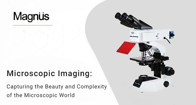
Microscopic Imaging: Capturing the Beauty and Complexity of the Microscopic World
Microscopic imaging reveals the hidden world beyond our sight. It entails taking precise pictures of small details or structures that are beyond the vision of the human eye. At the microscale, scientists can investigate cells, bacteria, and materials using specialized microscopes and imaging techniques.
Microscopic imaging is essential in several disciplines, such as biology, medicine, materials science, and nanotechnology. It helps scientists to investigate cellular functions, identify illnesses and examine the characteristics of materials. The knowledge of the world around you is enhanced due to these amazing technological advancements, which provide humanity with new perspectives on the delicate beauty and complexity of the tiny world. You will learn a lot about Microscopic Imaging in this post:
Importance of Microscopy and imaging:
Unfolding the unknown:
Microscopic imaging is similar to a wonderful window that opens to expose the world's hidden treasures. It gives scientists a glimpse into the microscopic realms that are invisible to the human eye.
For example, it reveals the secrets and activities of living things through the captivating dance of their cells. It also shows the atomic configurations of materials, even at the minuscule nanoscale.
Microscopic imaging unveils a world of astounding detail and complexity, stimulating curiosity and expanding scientific understanding, much as a microscope might expose the exquisite patterns on a butterfly's wing.
Let you understand biology secrets:
Scientific investigations into life's secrets are made possible using microscopic biology imaging. For instance, It can streamline biological processes and disclose the structure of DNA.
For example, Researchers may observe individual molecules inside cells using fluorescence microscopy.
It will also offer insight into processes like protein production and the development of illness. Microscopic imaging provides better insights into the basic methods of life by seeing these complex biological mechanisms.
Applications of microscopy and imaging:
The biological sampling
Advanced imaging techniques allow researchers to see the structure of human and model organism tissues and biomaterials without causing damage.
In this regard, X-ray technology has been quite helpful, as it allows for the dissection of dense samples without blood or discomfort.
Developments have significantly improved the imaging of biological material in time-resolved electron microscopy, increasing the image's contrast and resolution.
Material development
Producers must comprehend the qualities of their materials, whether creating alloys or processing plastics.
Microscopy and imaging techniques can be of considerable use in determining a material's strength, ductility, and hardness, as well as its resistance to various environmental factors, including temperature, pressure, and corrosion.
This information can then inform the uses and objectives for which the material can be utilized
Electronic Devices
These days, computer chips and other electronic equipment are so sophisticated and complicated that even piercing their outer shell might permanently harm the item.
Nevertheless, peeping inside these gadgets might occasionally be crucial to deciphering problems and carrying out fixes.
Microscopy and imaging techniques can provide a visual representation of the device's interior without harming its exterior or internal components.
Fossils and other delicate substances
It is vitally important that the specimen is not damaged while analyzing delicate materials like fossils or any other sensitive element.
Elements such as bone material, matrix composites, and fiber-reinforced polymers frequently utilized in bioengineering should be carefully analyzed.
Handling the materials is not advised due to their extremely brittle nature. However, sensitive imaging techniques, like helium microscopy, can circumvent this issue by providing researchers with an in-depth picture of the object without damaging it.
Advancements In Microscopy and Imaging:
Nanoscale Imaging Methods
Recent advances in microscopy have enabled scientists to see structures and processes at the nanoscale with unprecedented resolution and detail.
Scientists can now view molecular movements and interactions in real time due to techniques like high-speed atomic force microscopy (AFM) and super-resolution microscopy, which have elevated imaging capabilities.
These developments are transforming disciplines, including molecular biology, nanotechnology, and materials science, by providing fresh perspectives on the basic components of matter and life.
Multiple-modal Imaging Methods
An effective method for conducting thorough and multidimensional analysis is combining different imaging modalities.
By integrating techniques such as electron microscopy, spectroscopy, and fluorescence microscopy, researchers can link structural, chemical, and functional information inside a single sample with high resolution
This comprehensive approach opens the door for transdisciplinary discoveries and advances by improving the comprehension of intricate biological systems, material properties, and environmental occurrences.
Imaging in Vivo and Live-Cell Systems
The capacity to observe biological processes in living things in real-time is revolutionary in disciplines like cell biology, neuroscience, and pharmacology.
Modern imaging methods, such as light sheet microscopy and intravital microscopy, allow for the noninvasive observation of dynamic events in living tissues and organisms.
These techniques offer important new understandings of disease mechanisms and treatment approaches by capturing dynamic cellular activities, signaling pathways, and medication interactions within their natural environment.
High-Speed and Automated Image Processing
High-throughput imaging technologies and automated analysis algorithms have gained importance in response to the growing need for large-scale and rapid data capture.
Automated microscopy systems outfitted with robotic sample handling and image analysis AI enabled software enable the rapid screening of thousands of samples and the quantification of intricate biological or material features.
This faster data capture rate makes it easier to identify novel biomarkers, medication candidates, and materials with desirable qualities, which also speeds up scientific discovery.
Correlational Microscopy Methods
Complementary imaging techniques are combined in correlation microscopy to bridge the gap between various imaging modalities and length scales.
By correlating data from imaging techniques such as electron microscopy and optical microscopy, researchers can precisely pinpoint and define structures of interest inside complicated samples.
This integrated approach enhances comprehension of complex systems and phenomena, making it easier to conduct in-depth analyses of biological specimens, material interfaces, and cellular ultrastructure.
Final Words:
Microscopic imaging helps to explain the workings of the invisible world, including complex materials and cellular systems.
It provides insights into the workings of life and the characteristics of materials, with applications ranging from biology to materials science and beyond.
Recent developments like multi-modal methods and nanoscale imaging expand our comprehension.
Microscopic imaging is revolutionizing research and opening up new paths for innovation and discovery because it can quickly evaluate large volumes of data and view live systems in real time.